


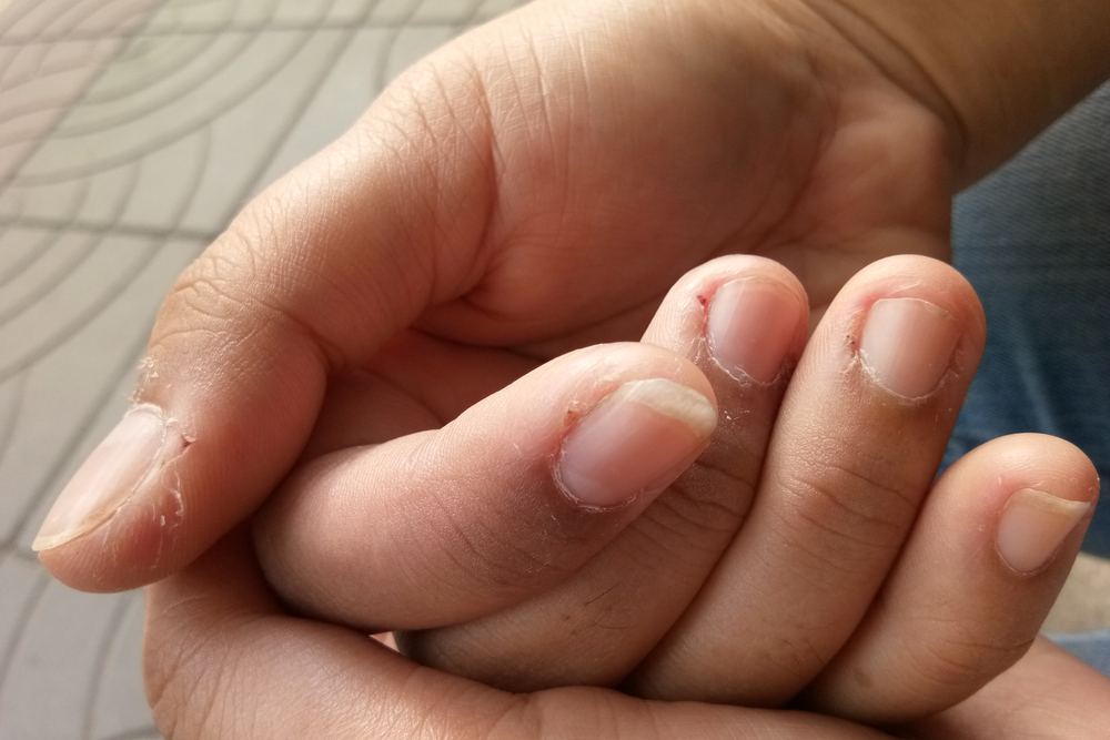
- Melanonychia is defined by a dark line on the fingernails or toenails.
- It can be the sign of a more serious illness, which is why getting a proper diagnosis is essential.
- A microscopic examination or biopsy is sometimes necessary to determine the cause and treatment options.
Finding the root cause of melanonychia is quite a complex process. Most cases are benign, but it can occasionally herald serious medical conditions. Here we describe the different types of melanonychia, how a diagnosis is made, and treatment options available to you.
What is melanonychia?
Melanonychia is a condition defined by a pigmented band within the fingernails or the toenails. It often only affects one or a few nails rather than all of them.
The unusually dark pigmentation is the result of melanin building up within the plate of the nail.
“Melanonychia is a Latin term. ‘Melano’ means black. In the human body, it refers to structures that are derived from melanocytes, which are the cells responsible for producing dark pigment. ‘Nychia’ refers to the nail,” explains Dr. Mohammed Ibrahim, Senior Research Fellow at Duke University in Durham, NC.
“Collectively, melanonychia is a condition caused by many disorders, all of which lead to dark discoloration of the nail.”
What causes dark lines on the nails?
Determining the cause of pigmentation in the nail is actually quite complicated. While typically harmless, the cases that have a potential health impact need to be identified and treated as soon as possible.
“Pregnancy, infections, malignancy — the list of the conditions that could cause melanonychia is long,” explains Ibrahim.
One of the most threatening conditions that need to be ruled out is melanoma — or skin cancer — within the nail. Every case of melanonychia should therefore be approached with concern in order to rule out any of the more malignant or life-threatening causes.
“Examining the nails can only confirm the presence of melanonychia, however, identifying the underlying cause is more important as this requires further systematic clinical investigation,” Ibrahim informs.
How is it diagnosed?
“When a patient presents signs of melanonychia, all twenty nails should be examined,” says Dr. Ibrahim.
He also recommends that the mucosal membranes of the nail be inspected for the presence of pigmented lesions. “The latter finding,” adds Dr. Ibrahim, “along with melanonychia are both associated with some systemic diseases, such as Addison disease.”
Next, the affected nails should be thoroughly examined using dermoscopy — a noninvasive inspection with a microscope.
“This test can be performed to diagnose fungal infections, but it also allows us differentiate between benign and malignant lesions,” Ibrahim explains. “A longitudinal melanonychia demonstrates dark brown lines or bands extending from the nail fold to the free edge of the nail, with a width less than 3 mm.”
Malignant nail melanomas, however, are characterized by irregular black, grey or brown lesions that involve more than two thirds of the nail. “These lesions may be associated with nail dystrophy,” Ibrahim notes.
Melanonychia in children
When a child presents with signs of melanonychia, even more questions arise.
For some, this could simply be part of their body’s growing pattern and the lines could easily disappear overtime. The majority of melanonychia in children simply mark the presence of a melanocytic nevus or mole under the nail.
While most cases prove to be benign, further testing is needed to confidently rule out the rare occurrence of melanoma.
The greatest concern and unanswered question is whether that benign pigmentation can become malignant later in life.
What if it’s cancer?
If the dermoscopy results suggest anything remotely malignant, the next step is a nail biopsy to confirm if there is melanoma within the nail. The most suspicious dermoscopic feature of melanoma in the nail is irregular lines on a brown background.
Other visual cues of melanoma are micro-Hutchinson’s sign — a wide pigmented band and triangular pigmentation on the nail plate.
There is currently no set evidence-based standard for the frequency of dermoscopic follow up, although most experts recommend checking suspicious longitudinal melanonychia every six months.
Patients with benign melanonychia are advised to keep a close eye on their nails and to report any developments or changes in the coloring or shape to their doctor immediately.
What are the common types of melanonychia?
There are three main types of melanonychia. All should be examined and watched closely to detect any changes over time.
Longitudinal melanonychia
This form of melanonychia is the most common and most harmless, according to a scientific review of the condition.
It can be the result of something as simple as a mole or freckle developing within the plate of the nail and producing a strange dark brown or black band down the middle of the nail.
Melanoma-related melanonychia
This form is hard to differentiate from an external exam alone, which is why patients with this type of melanonychia are often initially misdiagnosed.
Due to the delay in accurate diagnosis of this form of melanoma, which can stretch on for an average of two years, it carries a poor prognosis for patients.
Fungal melanonychia
This form of the disease may sometimes appear with white or yellow streaks but on the whole, it is still hard to differentiate from other forms.
Researchers have found that fungal melanonychia (FM) often forms a non-longitudinal pattern on the nail and tends to appear more blotchy in shape.
These visual cues alone, however, can not be used for diagnosis as FM has also appeared in gray colors, longitudinal patterns and other characteristics usually associated with melanoma. Thus, dermoscopy is the definitive test to properly diagnose FM.
How is melanonychia treated?
“The management of melanonychia depends on the type of lesion,” explains Dr. Ibrahim. “Benign longitudinal melanonychia usually require no further management, although the patient should periodically observe the lesion and seek medical advice if the pigmentation changes in shape, color, size, or if it starts bleeding.”
In the most severe cases, the treatment could be far more life-changing, notes Dr. Ibrahim. “Malignant lesions such as melanoma require aggressive management that may include amputation of part of the affected digit.”
He goes on to note that fungal melanonychia identified through dermoscopy should be treated using proper antifungal medications that corresponds to the fungus type.
» If you have dark lines on your nails, meet our medical review team for suggestions.